
Shoulder
1/15/2018
Scanning parameters are same as above.
*Arm in neutral position and built up so that elbow is level with shoulder joint.
*Axial plane along axis of humeral shaft.
*Coronal plane is from axials, parallel to the supraspinatus tendon/perpendicular to the plane in which the glenoid fossa articulates with the humeral head.
*Sagittal is perpendicular to the supraspinatus tendon/parallel to the plane in which the glenoid fossa articulates with the humeral head.
Arthrex Shoulder
12/08/2021
Scan supine, unaffected arm up over head, affected arm down by side; shoulder centered in gantry. Cover entire scapula and proximal 1/3 of humerus, FOV as small as possible, to include all of this.
Pitch of less than 1
No gantry tilt
140 kVp if available, otherwise 120 kVp
300 mAs with dose modulation: if modulation is not available, then 200 mAs or higher depending on patient size.
Helical 0.625 mm spacing x 0.625 mm thickness Standard Algorithm (1 X 1 on the Toshibas)
Recon 2: 0.625mm spacing x 1.25mm thickness Bone Algorithm
Recon 3: 2.5mm spacing x 2.5mm thickness Standard Algorithm
Sag/Cor 1.5 x 1.5mm Reformats from Recon 1 or 2 depending upon interest (1×1 on Toshibas)
Be sure to get axial, coronal, and sagittal between a combination of the initial acquisition and the Sag/Cor Reformats.
Send Initial acquisition, Recon 2, Recon 3, 3D, and Sag and Cor reformats

Shoulder DJO/Match Point
2/07/2020
Helical 0.625 mm spacing x 1.25 mm thickness Standard Algorithm (0.5 X 1 on the Toshibas)
Recon 2: 0.625mm spacing x 1.25mm thickness Bone Algorithm
Recon 3: 2.5mm spacing x 2.5mm thickness Standard Algorithm
Sag/Cor 1.5 x 1.5mm Reformats from Recon 1 or 2 depending upon interest (1×1 on Toshibas)
Be sure to get axial, coronal, and sagittal between a combination of the initial acquisition and the Sag/Cor Reformats.
Send Initial acquisition, Recon 2, Recon 3, 3D, and Sag and Cor reformats
Make a disc of ONLY first acquisition (i.e. Axial 0.625 x 1.25 Standard Algorithm, no edge enhancement, no tilt, no 3D, no reconstructions, no reformats, no lossy compression, primary DICOM images, at a pitch of 1), and send it with patient to Dr. Sanders’ office.
Patient Preparation
Remove any non-fixed metal prosthesis, jewelry, zippers that might interfere with the scan region to be scanned.
Make the patient comfortable and instruct him/her not to move during the procedure.
Position the patient as follows: supine, arms at sides of the body and with the shoulder in neutral rotation.
Cervical spine is in neutral position.
Place a marker on the arm indicating if it is the right or left. Use a marker that does not hinder the quality of the CT scan.
Scanning Instructions
Table position
Set the table height so that the area to be scanned is centered in the scan field.
Do not change table position between images so that all images create one unified volume. Field of view (FOV)
Capture the scapula and proximal humeral head using a 25cm FOV – DO NOT EXCEED.
Scan all slices with the same FOV, reconstruction center AND table height (coordinate system).
The scan range should cover the entire scapula. You may add axial slices as necessary to ensure coverage from superior to inferior.
Imaging soft tissue is unnecessary, only the bony regions are of interest.
No Gantry Tilt / No Oblique Images
Kernel
Use a soft tissue/moderate reconstruction algorithm, with no edge enhancement.
For DJO/Match Point:
Scan helical
Region of interest: Shoulder: full scapula and proximal humerus
Kv: 120
mAs: As given by the automatic system
Pitch: 1
Matrix: 512 x 512
FOV: 25cm x 25 cm – DO NOT EXCEED
Collimation:
Slice thickness: ≤ 1.25 mm
Slice increment: ≤ 0.625 mm (50% overlap)
Kernel / Algorithm: Moderate/soft tissue
Reconstruction and Delivery of the Images
No secondary reconstructions; images must be scanned at the given parameters or smaller.
No obliqueness; no gantry tilt and no oblique reconstructions.
No reformatting into coronal or sagittal planes; no MPR’s and no 3D reconstructions.
Provide the complete data set of primary DICOM images.
Lossy compression is NOT acceptable (ISO_10918_1, ISO_14495_1, ISO_15444_1 or ISO_13818_1).
Please include patient age, sex, and weight information.
Please retain a permanent archive (PACS) copy of the RAW data of images for 1 month.
Frequently Asked Questions
What should I do if the entire scapula doesn’t fit
in the 25cm FOV? It is not necessary to include the surrounding soft tissues, chest/lung or the clavicle in the FOV. You may add axial slices as necessary to increase coverage from superior to inferior.
What is the difference between bone kernel and soft tissue kernel? Bone kernel vs. soft tissue kernel: we want the soft tissue kernel with no edge detection because it is easier to process the 3D model from. The bone kernel is ideal for detecting fractures but this is not the purpose of these images.
Can we use compressed jpeg format? No,
DICOM images are necessary and the compressed jpeg format cannot be used to keep the accuracy of the 3D models. For more information, please contact Materialise at djoservice@materialise.be or 734-259-7014.
Match Point System DJO CT Scan Protocol Rev D Shoulder
https://djo.appdataroom.com/download/v/2251c3192df4044a6f4c9e85a3624a81cf152b6d405259?trackingGuid=f0795d6c-07cc-11ea-9be7-22000a99bb0a&groupId=8235

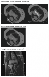
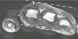
Elbow
Wrist

Hand/Finger
Orient coronal plane parallel to the volar (palmar)metacarpal head
Orient sagittal plane perpendicular to the coronal plane

Thumb
Orient axial plane perpendicular to midshaft of proximal thumb phalanx

Orient coronal plane parallel to thumb sesamoid bones, scan through entire thumb


Orient sagittal plane perpendicular to coronal plane, scan through entire thumb
